


Brunel Microscopes Ltd
Registered in U.K. (England) No: 2060047
find us on youtube, facebook and twitter


Vision Mantis Inspectoscope
Specialised Microscopes

Haemascope Dark Ground

The Haemascope is based on a research standard 100 watt high quality microscope that can be used for dark ground microscopy. It combines excellent performance with excellent value. For dark ground work, the Haemascope uses a high end oil immersion objective with an internal iris (to stop flare) and and oil immersion dark ground condenser developed specifically for this purpose..
The analysis of whole blood preparations has obtained considerable popularity as a diagnostic and health monitoring tool in a wide range of alternative medicine fields. Dark ground and phase contrast techniques are commonly used. However blood cells are poor reflectors of light and have little by way of internal structure and therefore require very effective microscopy to obtain the the images that are needed. Standard biological microscopes are not good enough for this application. We have to stress this point as too often we find that referrals to us have bought microscopes that cannot produce the images needed.
There are several units on the market that purport to offer the solution, and some are more effective than others. In essence the system requires two essential elements. Firstly an effective illumination system of 100 watts quartz halogen (LED illumination is not suitable) and secondly top end quality optics. The correct balance between these two factors is essential to obtain the results needed to apply these techniques professionally and safely. It is not possible to purchase a suitable system cheaply, they just won’t do the job.
Illumination
This is particularly important for dark ground analysis, which requires maximum illumination to overcome the fact that red blood cells are poor reflectors of light. A minimum of 100 watts is required for the light source, this is adequate providing the x100 objective is of good quality. The Haemascope has an excellent 100 watt quartz halogen light source. Some other units attempt to overcome poorer quality objectives by introducing excessive light by using fibre optic illumination introduced into the condenser of the microscope. This usually causes flare (which produces images that are unreal) and also an uneven field illumination, and generally tries to mask the fact that the x100 objective of the system is just not of sufficient quality for the purpose.
Objectives
The objectives of any microscope system are the essential components necessary to achieve the results required. Failure to address this fact by attempting to overcompensate with unnecessary over illumination is a significant criticism of some units. For dark ground analysis, the Haemascope incorporates a high quality plan x100 objectives with an internal iris diaphragm.
Other Factors
Attention to detail is all important, and the types of slides, coverslips and even immersion oil can have a significant effect. Brunel Microscopes supply, with both its recommended systems, the best quality slides and coverslips of the correct thickness and our own formulated immersion oil.
Brunel Haemascope
Live cell analysis of whole blood from a finger prick site involves dark ground microscopy. . The unit comprises:
Trinocular microscope with x2.5 x4, x10, x40 and x100 (with iris diaphragm) objectives.
High specification oil darkground condenser
100 watt quartz halogen illumination system
The key to successful darkground analysis of whole blood rests with the illumination system and the quality of the x100 oil objective. The high wattage halogen Kohler illumination of the Haemascope has been developed specifically for this purpose, as has the x100 objective which is an excellent lens with an internal iris diaphragm. The two combine to give images that are the equal of units costing many times the price.
The Haemascope is preset to minimise the need for setting up the optics, simply make the slide preparation and follow a few basic instructions for instant results.
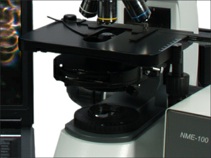

Brunel Haemascope mechanical stage
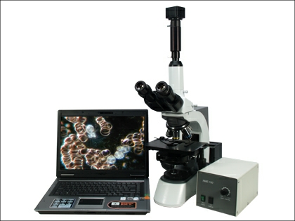
Brunel Haemascope Microscope
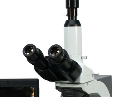
Brunel Haemascope Trinocular head
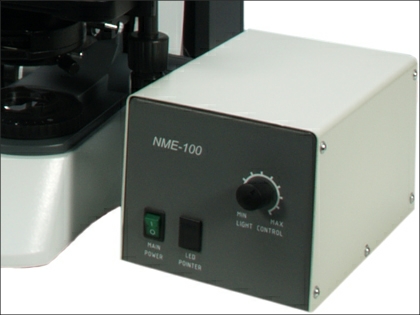
Brunel Haemascope illumination control box
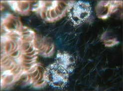
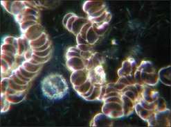
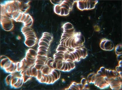
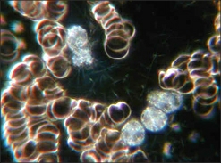
Dark ground images


| Pocket microscopes /hand lenses |
| Budget Stereos |
| Lab Stereos |
| Zoom Stereos |
| Long arm Stereos |
| Gemscopes |
| Inspection & QC Stereos |
| Zoom Monoscopes |
| High Power Digital |
| Handheld & Desk Top |
| Digital Stereos |
| Digital Cameras |