


Disease Identification
There are basically two types of microscope, those that are high power for looking at slide preparations at things that are microscopic (can magnify x40 up to x1000), and those that are low power stereomicroscopes (usually magnifying between x10 and x40 or so) and are used for looking at whole objects and are ideal for bee dissection. Unfortunately because of the laws of physics that govern light it is not possible to have a microscope that can do both functions. We are able to offer budget equipment at a realistic price for the individual and also more sophisticated instruments more suited to the needs of societies and education.
Diseases that can be identified with a high power microscope
Bee diseases are the result of either poor apiculture technique, bacteria, fungus, parasite infestation or virus infection.
Viruses (for example Sacbrood) cannot be seen with a light microscope.
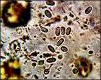
Bacteria can be seen at magnifications of x1000. However there are only two physical shapes that bacteria can have, either ‘sausage’ shape -
Recently published and recommended books
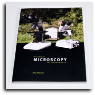
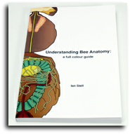
Both these recently published books make very useful reference works and cover between them the detailed structure of the bee and provides a very practical guide to using microscopes in beekeeping.
The author, Bob Maurer, has acquired considerable knowledge in this area from a self taught beginning. This book is well written from this perspective and is an easy read. An excellent book.
Price £15.00
Understanding honeybee anatomy is a magnificent book with a very large number of full colour photographs that explain visually the structure of the honeybee. The modern definitive work by Ian Stell.
Price £27.50
Fungus (Chalk and Stone Brood) is much easier to identify with a high power microscope and well within the abilities of the beginner.
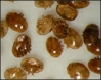
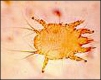
Parasites (Varroa mite, Nosema, Acarine and Amoeba) are again easy to see with a microscope. The varroa mite can be seen with a low power stereomicroscope because of its relatively large size, and the trachea of the bee needs to be dissected out to make a slide for high power examination to identify acarine and the ventriculus for nosema.
Pollen Identification
The identification of the principal pollen content of honey can be a commercial necessity for those producers who wish to feature this as a selling point. It also adds considerable interest for the general honey producer to track the crops preferred by their hives. Little specialist equipment is needed for this and we provide a Pollen Identification Kit to make this easy . This includes the book ‘Pollen its identification and preparation for the microscope’ by J. White. Kit contents include specimen tubes, watch glasses, pipettes, ringing brush, isopropyl alcohol, ringing cement, glycerine jelly for pollen, pollen identification by White, slides, coverslips, and instructions. Click here for Pollen Slide Preparation.
Price -
Artificial Insemination
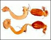
Artificial insemination of the queen is now a well established technique with a number of insemination devices on the market. This is a two step process starting with the inversion of the drone to collect semen, then the careful insemination using the precision manipulation units that immobilise the queen and allows introduction of the semen.
This is a delicate and precise operation and it is essential to use a low power stereomicroscope, either one with a large area base to take the inseminator or a long arm version that allows the stereohead to be held above the machine.

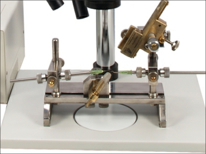

Insemination stage area
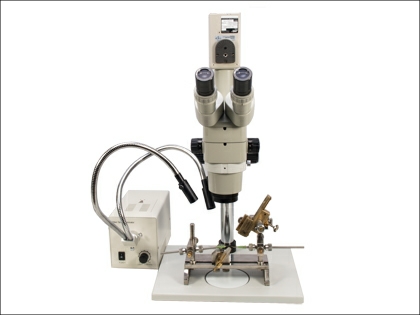
Brunel Bee Artificial Insemination microscope


Brunel Microscopes Ltd
Registered in U.K. (England) No: 2060047
find us on youtube, facebook and twitter


Beekeeping and microscopy explained
Beekeeping


 Secure Shop
| Pocket microscopes /hand lenses |
| Budget Stereos |
| Lab Stereos |
| Zoom Stereos |
| Long arm Stereos |
| Gemscopes |
| Inspection & QC Stereos |
| Zoom Monoscopes |
| High Power Digital |
| Handheld & Desk Top |
| Digital Stereos |
| Digital Cameras |